
Introduction
Lipid nanoparticles (LNPs) have rapidly emerged as the leading non-viral delivery platform for nucleic acid therapeutics, underscored by the clinical and commercial success of mRNA-based COVID-19 vaccines. Unlike conventional small-molecule drugs, LNP-formulated therapeutics present unique analytical challenges: their performance depends not on a single molecular entity but on a constellation of interdependent physicochemical properties. Particle size, morphology, surface charge, and lipid crystallinity collectively govern critical behaviors — from in vivo biodistribution and cellular uptake to payload release kinetics and long-term storage stability.
For research teams advancing nucleic acid drug candidates through preclinical development, robust characterization is not merely a quality control checkbox — it is fundamental to de-risking programs and building the data packages that regulators expect. Comprehensive Lipid Nanopartical-formulated Drug Characterization encompasses a suite of orthogonal analytical techniques that together establish the critical quality attributes (CQAs) defining an LNP product’s identity, purity, and performance. This article examines the four foundational pillars of LNP characterization — particle size, morphology/ultrastructure, surface charge, and crystallinity/polymorphism — and discusses how an integrated analytical strategy can accelerate the translation of nucleic acid nanomedicines toward the clinic.
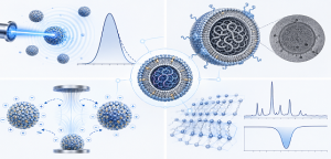
The Importance of Multimodal LNP Characterization
A defining feature of LNPs is that their four core physical properties do not operate in isolation. Changes in one CQA frequently cascade into others. For instance, lipid oxidation during storage at 25 °C may go undetected by standard particle sizing yet can subtly alter surface charge distribution and promote polymorphic transitions in the lipid matrix — changes that collectively compromise encapsulation efficiency and therapeutic potency. This interdependence demands that characterization strategies employ complementary, orthogonal methods rather than relying on any single analytical readout.
Regulators increasingly expect sponsors to define CQAs using a Quality-by-Design (QbD) framework, wherein each physicochemical attribute is linked to a functional outcome. For example, the mean hydrodynamic diameter measured by dynamic light scattering (DLS) may serve as a surrogate for systemic circulation half-life, while the zeta potential threshold required for colloidal stability (typically |±30 mV|) can be validated against aggregation kinetics under accelerated storage conditions. This mechanistic understanding transforms characterization data from a retrospective QC metric into a prospective development tool.
Particle Size and Polydispersity: Gatekeepers of Biodistribution
Among all LNP CQAs, particle size arguably exerts the most direct influence on biological fate. Nanoparticles in the 100–300 nm range are generally favored for systemic delivery: this size window balances sufficient circulation time to reach target tissues against the risk of rapid hepatic clearance. Particles below approximately 10 nm undergo swift renal filtration, while those exceeding 300 nm are preferentially sequestered by the mononuclear phagocyte system in the liver and spleen. LNP Particle Size Characterization provides not only the mean hydrodynamic diameter (Z-average) but also the polydispersity index (PDI) — a dimensionless measure of distribution width that is frequently the first indicator of emerging instability.
DLS remains the workhorse technique for routine sizing, valued for its speed, minimal sample requirements, and broad compatibility with aqueous suspensions. However, DLS has well-characterized limitations: it is intensity-weighted and therefore disproportionately sensitive to larger particles or aggregates, and it cannot resolve multimodality in heterogeneous populations. Nanoparticle tracking analysis (NTA) complements DLS by tracking the Brownian motion of individual particles, providing number-weighted size distributions and direct particle concentration measurements. For the most rigorous characterization, asymmetrical flow field-flow fractionation (AF4) coupled with multi-angle light scattering can physically separate subpopulations prior to analysis, resolving subtle size shifts that DLS alone would average into obscurity.
A particularly instructive finding from recent benchmarking studies is that LNP size does not appear to be strongly influenced by RNA cargo length — a counterintuitive observation that underscores the importance of empirical characterization rather than relying on theoretical predictions. For development teams, adopting a multi-instrument sizing strategy — DLS for routine monitoring, NTA for concentration and subpopulation analysis, and AF4 for high-resolution separation — provides the orthogonal data needed for robust CMC documentation.
Morphology and Ultrastructure: Seeing Is Believing
While particle sizing techniques provide essential statistical descriptors of LNP populations, they reveal nothing about individual particle structure. Morphology & Ultrastructure Characterization fills this gap by providing direct visual evidence of particle shape, lamellarity, internal organization, and the presence — or absence — of structural defects. For nucleic acid-loaded LNPs, this information is critical: a uniformly sized population visible by DLS may nevertheless contain a heterogeneous mix of properly encapsulated particles, empty liposomes, and fused aggregates, each with profoundly different biological activity.
Cryogenic transmission electron microscopy (cryo-TEM) is widely regarded as the gold standard for LNP ultrastructural analysis. By rapidly vitrifying samples in liquid ethane at approximately −196 °C, cryo-TEM preserves particles in a near-native hydrated state, avoiding the dehydration and staining artifacts that can distort lipid bilayers in conventional TEM preparations. At resolutions approaching 0.1–1 nm, cryo-TEM can resolve individual lipid bilayer leaflets, distinguish unilamellar from multilamellar vesicles, and visualize the electron-dense nucleic acid core that confirms successful payload encapsulation.
For applications requiring complementary surface information, atomic force microscopy (AFM) provides three-dimensional topographical data — height, width, and surface roughness — that can reveal subtle morphological changes induced by environmental factors such as pH shifts or temperature fluctuations. Scanning electron microscopy (SEM), while less commonly used for sub-100 nm LNPs, offers valuable three-dimensional perspective on larger aggregates and can confirm overall population homogeneity. The choice among these techniques should be guided by the specific question at hand: cryo-TEM for internal structure, AFM for surface topography, and SEM for aggregate characterization.
Surface Charge: The Electrostatic Determinant of Stability and Interaction
The surface charge of an LNP, most commonly reported as zeta potential (ZP), is a primary determinant of colloidal stability. Particles with ZP magnitudes exceeding approximately ±30 mV generate sufficient electrostatic repulsion to resist aggregation, while values approaching zero permit van der Waals forces to drive particle coalescence. Beyond stability, surface charge profoundly influences interactions with biological environments: strongly cationic particles may exhibit efficient cellular uptake but risk non-specific binding to serum proteins and associated toxicity, whereas near-neutral surfaces generally demonstrate improved systemic compatibility.
Surface Charge Characterization extends beyond a single ZP measurement to encompass surface charge density, isoelectric point (pI) determination, and charge uniformity across the particle population. Laser Doppler electrophoresis (LDE) provides rapid, routine ZP measurements from microliter-scale sample volumes. Potentiometric titration offers deeper insight by mapping ZP as a function of pH, identifying the optimal buffer conditions for long-term stability and the pI at which aggregation risk is maximized.
A critical but often underappreciated dimension of surface analysis is the characterization of the biomolecular corona — the layer of serum proteins that spontaneously adsorbs onto LNP surfaces upon systemic administration. This protein coating, rather than the pristine nanoparticle surface, is what cells and tissues actually encounter. Recent work using surface plasmon resonance (SPR) has demonstrated that measuring LNP affinity for key plasma proteins such as apolipoprotein E (ApoE) can provide early, predictive insight into organ tropism — high ApoE binding, for example, strongly correlates with hepatocyte targeting via the low-density lipoprotein receptor pathway. Integrating SPR-based corona analysis with conventional ZP measurements creates a more complete picture of how an LNP formulation will behave in the complex in vivo environment.
Crystallinity and Polymorphism: The Solid-State Foundation of LNP Performance
Beneath the colloidal surface of LNPs lies a lipid matrix whose solid-state organization — degree of crystallinity, polymorphic form, and phase behavior — fundamentally governs drug loading capacity, release kinetics, and long-term physical stability. Crystallinity & Polymorphism Characterization employs a suite of analytical techniques to elucidate these hidden structural features.
Triglyceride-based lipids, common in LNP formulations, are particularly susceptible to polymorphic transitions during processing and storage. The least stable α-form, characterized by a hexagonal subcell packing, can transition to the more ordered β′-form and ultimately to the thermodynamically stable β-form with triclinic packing. Each transition is accompanied by increasing lipid packing density, which reduces the amorphous domains available for drug accommodation — a process that may progressively expel encapsulated payload and contribute to the shelf-life limitations observed in many early-stage LNP formulations.
Differential scanning calorimetry (DSC) is the primary tool for characterizing these thermal transitions, detecting endothermic melting events and exothermic recrystallization peaks that reveal the polymorphic landscape of the lipid matrix. X-ray diffraction (XRD) provides complementary crystallographic information at the atomic scale, with characteristic short-spacing peaks that unambiguously identify polymorphic forms: the α-form at approximately 4.15 Å, the β′-form as a doublet at 3.8 and 4.2 Å, and the β-form with a strong reflection at 4.6 Å. Small-angle X-ray scattering (SAXS) extends this analysis to the mesoscale, revealing the long-range ordering of lipid bilayers and enabling the identification of non-lamellar phases — notably the inverse hexagonal (HII) phase — that are associated with enhanced endosomal escape and transfection efficiency.
For development programs, establishing a comprehensive thermal and crystallographic profile early in formulation development helps anticipate stability liabilities before they manifest in costly late-stage failures. The QbD principle applies here as well: by defining the acceptable polymorphic window — the range of crystalline states within which product performance remains consistent — teams can establish meaningful specifications for manufacturing control and stability monitoring.
An Integrated Analytical Strategy for Preclinical Development
The four CQA domains discussed above — size, morphology, surface charge, and crystallinity — are not independent variables. An effective characterization strategy recognizes their interdependence and employs orthogonal techniques that cross-validate one another. For example, a batch that passes routine DLS-based size specifications may harbor subtle morphological defects detectable only by cryo-TEM. Conversely, a formulation with an acceptable zeta potential profile may undergo undetected polymorphic transitions during long-term storage that progressively compromise encapsulation efficiency.
For research teams working at the forefront of nucleic acid therapeutics, the analytical burden can be substantial. Each characterization modality requires specialized instrumentation, method development expertise, and experience interpreting the resulting data in the context of specific therapeutic applications. Partnering with an experienced preclinical CRO can provide access to a comprehensive analytical toolkit — DLS, NTA, AF4, cryo-TEM, AFM, SEM, LDE, SPR, DSC, XRD, and SAXS — without the capital investment and personnel costs of building these capabilities in-house. Moreover, an integrated outsourcing model ensures that data from different techniques are interpreted holistically, identifying correlations between physicochemical parameters and functional outcomes that might be missed when each analysis is performed in isolation.
Conclusion
Robust lipid nanoparticle characterization is a prerequisite, not an afterthought, for successful nucleic acid therapeutic development. The four pillars of CQA analysis — particle size and distribution, morphology and ultrastructure, surface charge and biomolecular interactions, and crystallinity and polymorphism — together define the identity, stability, and predict the biological performance of LNP-formulated drugs. Investing in comprehensive, orthogonal characterization early in development yields dividends throughout the product lifecycle: it accelerates formulation optimization, strengthens regulatory submissions, and reduces the risk of late-stage surprises.
For research groups seeking to navigate the analytical complexity of LNP development, Creative Biolabs offers integrated preclinical CRO services spanning the full characterization continuum. From initial formulation screening through IND-enabling stability studies, our scientific team works collaboratively with clients to design tailored analytical strategies that align with specific program objectives and regulatory milestones. To discuss how a customized characterization plan can support your nucleic acid therapeutic program, contact our team to explore project-specific solutions.
FAQ
Q: What are the critical quality attributes (CQAs) for lipid nanoparticle-formulated drugs?
A: The primary CQAs for LNPs include particle size and polydispersity index (PDI), zeta potential (surface charge), morphology and ultrastructure, encapsulation efficiency, lipid crystallinity and polymorphism, and colloidal stability under relevant storage and physiological conditions. Each CQA is linked to a specific aspect of product performance — for instance, size influences biodistribution, while zeta potential governs colloidal stability and cellular interactions.
Q: Why is cryo-TEM preferred over conventional TEM for LNP characterization?
A: Cryo-TEM preserves LNPs in a near-native hydrated state through rapid vitrification at cryogenic temperatures, avoiding the dehydration, chemical fixation, and heavy-metal staining required for conventional TEM. These preparative artifacts can distort fragile lipid bilayers, collapse internal structures, and obscure the electron-dense nucleic acid core. For LNP-mRNA systems especially, cryo-TEM is strongly recommended to obtain accurate ultrastructural information.
Q: How does lipid polymorphism affect LNP stability and drug release?
A: Lipid polymorphism refers to the ability of lipid molecules to adopt different crystalline packing arrangements. During processing and storage, triglycerides in LNP formulations may transition from the less stable α-form through the β′-form to the most stable β-form. Each transition increases lipid packing density and reduces the amorphous volume available for drug accommodation, which can progressively expel encapsulated payload and contribute to shelf-life limitations. Characterizing and controlling polymorphic behavior through DSC and XRD is therefore essential for ensuring consistent product performance.
Q: What is the significance of the biomolecular corona in LNP surface characterization?
A: Upon systemic administration, serum proteins rapidly adsorb onto LNP surfaces, forming a biomolecular corona that effectively becomes the interface encountered by cells and tissues. This protein coating — rather than the pristine nanoparticle surface — determines opsonization, clearance kinetics, and receptor-mediated targeting. Characterizing the corona through techniques such as SPR-based affinity measurements for key proteins like ApoE provides early, predictive insight into organ tropism and can help de-risk preclinical translation.
Q: How can partnering with a preclinical CRO support LNP characterization programs?
A: An experienced preclinical CRO can provide access to a comprehensive suite of orthogonal analytical techniques — including DLS, NTA, cryo-TEM, AFM, DSC, XRD, SAXS, and SPR — without the capital investment and specialized personnel required to build these capabilities in-house. Beyond instrumentation, CRO partnerships offer method development expertise, QbD-aligned study design, and regulatory-grade reporting that accelerates IND-enabling data package preparation.
References
- Hallan, Supandeep Singh, et al. “Challenges in the physical characterization of lipid nanoparticles.” Pharmaceutics13.4 (2021): 549. CC BY 4.0. https://doi.org/10.3390/pharmaceutics13040549